AI-Powered TP53 Mutation Prediction: A Pan-Cancer Computational Pathology Study
A new AI model analyzes routine H&E pathology slides to predict TP53 mutations, RNA expression, tumor type and survival outcomes across 32 cancer types.
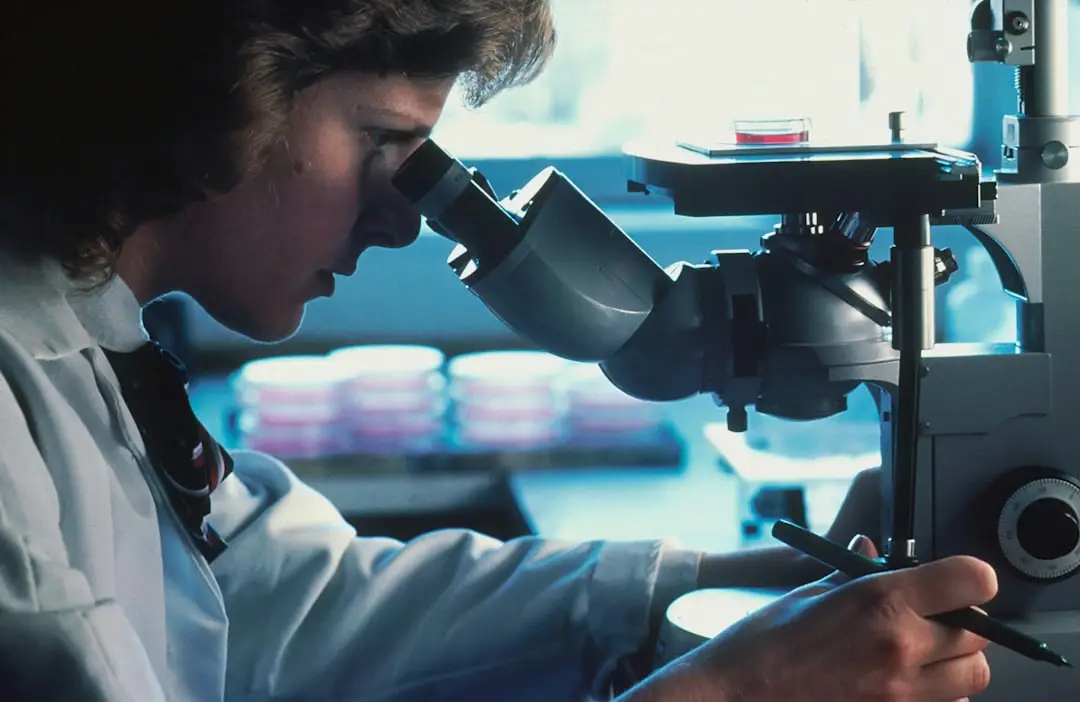
AI Could Extract Molecular Clues From Routine Cancer Pathology Slides
Artificial intelligence is opening a new avenue for computational pathology and precision oncology.
Researchers have developed a multitask AI model capable of analyzing routine hematoxylin and eosin (H&E)-stained whole-slide pathology images and extracting information that traditionally requires additional molecular testing.
The model was designed to simultaneously predict TP53 mutation status, TP53 RNA expression, tumor type and survival-related outcomes across 32 types of solid cancer.
The research, published in The American Journal of Pathology, demonstrates how AI could potentially connect what pathologists see under the microscope with the molecular characteristics of a tumor.
Importantly, the technology is being investigated as a decision-support and screening tool, rather than a replacement for established molecular testing.
Why TP53 Matters in Cancer
The TP53 gene is one of the most frequently altered tumor-suppressor genes in human cancers.
The protein produced by TP53 plays an important role in regulating cell growth and responding to cellular damage. When TP53 is altered, cancer cells can acquire characteristics that contribute to tumor progression and treatment resistance.
Determining TP53 mutation status normally requires molecular or genomic testing.
However, access to comprehensive molecular profiling can vary between healthcare systems, particularly in resource-limited or remote settings.
This has created interest in whether routinely collected pathology images could provide additional information about a tumor’s molecular biology.
How the AI Model Works
The researchers developed a Vision Transformer (ViT)-based artificial intelligence model combined with a multiple-instance learning approach.
Instead of requiring a separate AI system for every prediction, the model was designed as a multitask system capable of producing several outputs from the same whole-slide image.
The researchers used more than 11,000 primary tumor cases from the Pan-Cancer Atlas, together with corresponding:
- Somatic mutation data
- RNA-sequencing data
- Clinical information
- Survival outcomes
- Histopathology whole-slide images
The pathology images underwent preprocessing that included tissue masking, quality control, stain normalization, patch extraction and feature embedding before being analyzed by the AI system.
This approach is significant because whole-slide pathology images are extremely large and contain millions of visual features.
AI Predicts TP53 Mutations Across 32 Cancer Types
One of the most notable findings was the model’s ability to predict TP53 mutation status across 32 different solid tumor types.
When evaluated on an independent validation dataset containing 1,729 whole-slide images, the model achieved an overall AUROC of 0.766 for TP53 mutation detection.
The reported balanced accuracy was approximately 0.72, with sensitivity of 0.70 and specificity of 0.73.
The model also demonstrated strong performance in identifying tumor types, with AUROC values above 0.88 for most tumor classes in the unseen dataset, although ovarian tumors were an exception.
These results suggest that microscopic tissue architecture may contain patterns associated with underlying molecular alterations that can be detected computationally.
From Histology Images to Molecular Biomarkers
The broader concept behind this research is sometimes described as image-based molecular profiling.
Traditionally, pathology and molecular diagnostics operate as complementary disciplines:
Pathology → What does the tumor look like?
Genomics → What molecular alterations does the tumor contain?
AI potentially provides a bridge between these two layers.
By learning relationships between histological patterns and molecular data, computational pathology models can attempt to infer biomarkers directly from tissue images.
Previous pan-cancer research has demonstrated the broader potential of this approach. A 2024 study trained more than 12,000 deep-learning models covering 4,031 biomarkers across 32 cancer types, including mutations, transcriptomic markers, protein expression, molecular subtypes and clinical outcomes.
The new TP53 study takes a different approach by bringing several prediction tasks together within a single multitask model.
Why This Could Matter for Precision Oncology
The potential clinical value is particularly interesting in settings where comprehensive molecular testing is difficult to access.
An AI system that analyzes an existing pathology slide could potentially help clinicians:
- Identify patients who may benefit from additional molecular testing
- Prioritize samples for genomic analysis
- Support diagnostic workflows
- Provide additional information when molecular testing is delayed
- Assist pathologists in interpreting complex tumor characteristics
- Potentially improve access to biomarker-driven cancer care
Rather than replacing molecular diagnostics, the model could eventually function as a triage or decision-support layer.
Researchers emphasize that confirmatory molecular testing would still be necessary before using a biomarker prediction for clinical treatment decisions.
The Role of Weakly Supervised Learning
One of the technical challenges in computational pathology is the enormous size of whole-slide images.
Manually labeling every relevant tumor region requires substantial expert time and can introduce variability between observers.
The researchers therefore used a weakly supervised learning strategy.
Instead of requiring detailed annotations for every individual region of the slide, the model can learn from slide-level information and identify patterns associated with the target outcomes.
This approach is particularly useful for large pathology datasets because it reduces the need for exhaustive manual annotation.
What Makes This Approach Different?
Many existing AI systems in oncology are designed for a relatively narrow task.
For example:
Model A → Tumor classification
Model B → Mutation prediction
Model C → Survival prediction
The new approach attempts to combine multiple objectives into one architecture.
The model can simultaneously generate information related to:
- TP53 mutation status
- TP53 RNA expression
- Tumor classification
- Survival-related outcomes
- Additional pathology-derived predictions
This multitask design could potentially make AI systems more useful within real-world pathology workflows.
AI in Pathology: From Image Recognition to Biological Inference
The significance of this research extends beyond TP53.
Traditional digital pathology largely focuses on recognizing visual patterns—for example, identifying tumor cells, classifying tissue types or quantifying specific morphological features.
The emerging field of AI-powered computational pathology is moving toward a more ambitious objective:
Can microscopic tissue morphology reveal hidden information about tumor biology?
If the answer is yes, pathology images could become computationally interpretable sources of molecular information.
This could create new connections between:
Histology → Molecular biology → Biomarkers → Prognosis → Treatment decisions
Such an approach fits closely with the broader goals of precision oncology.
Important Limitations
Despite the promising results, this technology is not yet a substitute for genomic or molecular testing.
Several challenges remain before AI-based biomarker prediction can become routine clinical practice.
1. External validation
Models trained using large research datasets need to be tested across independent hospitals, laboratories, scanners and patient populations.
2. Image variability
Differences in staining protocols, tissue preparation, scanners and image quality can affect AI performance.
3. Biological complexity
A visual prediction does not necessarily establish the biological mechanism responsible for the observed association.
4. Clinical validation
A model must demonstrate that its predictions improve clinically meaningful outcomes before it can be incorporated into routine decision-making.
5. Molecular confirmation remains important
AI predictions should currently be viewed as complementary to established molecular diagnostic methods.
What This Means for the Future of AI in Oncology
The study illustrates an important direction for AI in cancer research.
Instead of treating pathology images as purely visual diagnostic material, researchers are increasingly exploring them as potential sources of molecular and prognostic information.
Future computational pathology systems could potentially integrate:
- Histopathology
- Genomics
- Transcriptomics
- Proteomics
- Clinical records
- Treatment response
- Patient outcomes
Such multimodal AI systems could provide a more comprehensive representation of tumor biology.
The ultimate goal would not be to replace oncologists, pathologists or molecular laboratories, but to give them more information from the same clinical data.
Key Takeaways
- Researchers developed a multitask AI model for computational pathology.
- The model analyzes routine H&E-stained whole-slide images.
- It can predict TP53 mutation status across 32 solid cancer types.
- The model achieved an overall AUROC of 0.766 for TP53 mutation prediction on an independent validation set.
- It can also infer TP53 RNA expression and classify tumor types.
- The training dataset included more than 11,000 primary tumor cases.
- The technology could potentially help prioritize patients for molecular testing.
- The approach could improve access to biomarker information in resource-limited settings.
- The AI system should currently be considered a screening or decision-support technology, not a replacement for molecular testing.
The Bigger Picture: Can a Pathology Slide Become a Molecular Test?
The most exciting implication of this research may be the possibility that a routine pathology image contains more information about cancer biology than is immediately visible to the human eye.
AI can analyze thousands or millions of microscopic patterns simultaneously and search for relationships between tissue morphology and molecular characteristics.
If these models can be reliably validated across diverse clinical environments, routine pathology slides could eventually become an additional computational source of information for cancer biomarker assessment and precision medicine.
The future of oncology may therefore involve not only reading what a tumor looks like—but using AI to understand what its microscopic appearance may reveal about its underlying biology.
Source and Further Reading
The research was published as “Predicting TP53 Biomarkers from Whole Slide Images across Human Solid Tumors Using Weakly Supervised Learning” in The American Journal of Pathology.
Research DOI: 10.1016/j.ajpath.2026.05.008.
Medical Disclaimer
This article is intended for educational and informational purposes only. AI-based biomarker predictions are an emerging research technology and should not be used as a substitute for validated molecular testing, pathology interpretation or professional medical advice.
Educational content only. This material is written for healthcare professionals and students. It is not medical advice, and it must not be used for diagnosis or treatment decisions. Clinical decisions remain the responsibility of a qualified healthcare professional. Full disclaimer
Related resources

Merck and Moderna announced positive topline results from the Phase 3 INTerpath-001 trial for an individualized mRNA-based cancer therapy in melanoma.
Discover how AI-powered clinical trial matching can help breast cancer patients and oncologists identify potentially relevant clinical trials faster—and understand its benefits and limitations.
GenBio AI's AIDO Cell aims to simulate human cells from DNA and RNA to proteins and whole-cell behavior. Explore what virtual cells could mean for drug…